What To Expect When Your Pet Has Cataract Surgery
Surgery

Cataracts are a common cause of vision impairment in dogs, and less commonly a cause of vision impairment in cats. You may be familiar with cataracts and cataract surgery for yourself or a family member. Cataract surgery in veterinary medicine has made many advances and is one of the most rewarding procedures we can perform with success rates in dogs reported from 85-95% in an uncomplicated case. In our patients, surgery has some similarities and some big differences to expect compared to surgery in people.
Continue reading to learn more about the Ophthalmology department at Virginia Veterinary Centers, and how our board-certified veterinary ophthalmologist can address your pet’s cataracts.
WHAT ARE CATARACTS AND WHEN SHOULD YOU CONSIDER SURGERY FOR YOUR PET?
A cataract is an opacity in the lens inside of the eye and can range from very small (incipient) to complete and blinding. At this time, there is no medical therapy that can reverse the changes to the lens once they have occurred. Cataracts in dogs are most commonly due to inherited reasons, followed by cataracts secondary to diabetes mellitus. Cats generally get cataracts secondary to chronic intraocular inflammation, though they can also have inherited cataracts.
The goal of surgery is to take a pet that is significantly visually impaired and restore vision, so we generally recommend surgery when cataracts have started to affect visual function. While cataract surgery is elective in veterinary patients, if left untreated, cataracts can cause secondary inflammation inside the eye that can sometimes lead to painful changes. Even if surgery is not an option, there are medications that can help control inflammation and prevent these complications, so an evaluation with a board-certified ophthalmologist may still be recommended.
WHAT TO EXPECT AT YOUR FIRST OPHTHALMOLOGY APPOINTMENT FOR YOUR PET
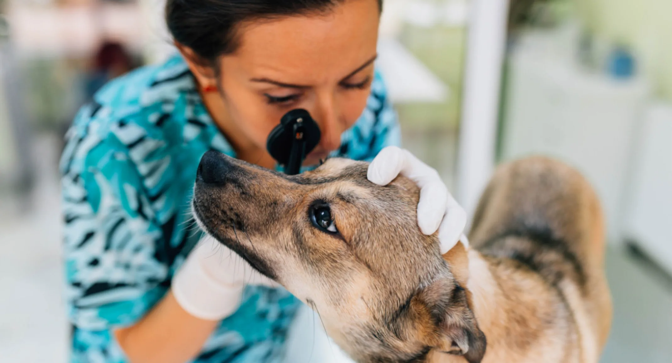
During your pet’s first visit to the Ophthalmology department, we will perform a complete ophthalmic examination including:
Testing tear production
Checking for corneal ulcers
Measuring intraocular pressure
We evaluate your pet’s overall ocular health and the progression of the cataract to help determine candidacy for surgery. It is also important to evaluate the health of the retina (the “film in the camera”), which we often cannot see through the cataract. This requires advanced diagnostics, including an electroretinogram to make sure the retina responds normally to light, and an ocular ultrasound to make sure the structure of the back of the eye is normal.
We will often start topical medications at your first appointment to prepare for surgery. We also discuss the process of surgery, success rates, and possible complications as they may apply to your pet.
OUR OPHTHALMOLOGY TEAM PARTNERS WITH YOUR PRIMARY CARE VETERINARIAN
Your primary care veterinarian is vital to your pet’s whole-body health and is a partner in their care. Because your pet has to undergo general anesthesia for cataract surgery, we recommend general bloodwork and urinalysis to help determine if there are any systemic concerns prior to surgery and anesthesia. These tests may be performed with your primary care veterinarian. Your veterinarian is also crucial in helping manage any other systemic concerns that could impact the outcome of surgery, such as diabetes mellitus, dental disease, skin infections, heart disease, etc.
WHAT HAPPENS DURING YOUR PET’S CATARACT SURGERY?
On the morning of your pet’s cataract surgery, your pet will need to be dropped off early to receive preoperative eye drops and for our team to make other preparations for their procedure. Your pet will be placed under general anesthesia for cataract surgery. We typically recommend doing surgery on both eyes, as long as both eyes are good candidates. In human cataract surgery, oftentimes surgery will be handled one eye at a time.
During cataract surgery, a very small incision is made in the cornea, then a circular hole is carefully torn in the lens capsule. The lens is broken up and removed with ultrasound energy (phacoemulsification) and an intraocular lens is placed in the lens capsule to focus light on the retina. Sometimes an artificial lens cannot be placed, resulting in far-sightedness in that eye. At the conclusion of surgery, the corneal incision is closed with very small sutures.
Most pets can return home in the evening the same day their cataract surgery takes place. They often benefit from relaxing at home to recover in their normal environment. If this is the case, the patient will return the following day for their first postoperative recheck. Fortunately, uncomplicated cataract surgeries are not painful procedures – most pets seem to experience only mild light sensitivity.
SHOULD I BE CONCERNED ABOUT MY PET’S CATARACT SURGERY?
Complications are possible with any procedure, and cataract surgery is no exception. The complication rate ranges from 5-15% and some patients may be at higher risk depending on the cause of their cataract, breed, age, and other factors. Some possible complications include:
Corneal Ulceration
– A break in the corneal surface layer; some ulcers are superficial and minor, and generally, heal easily with treatment. If left untreated, corneal ulcers may become infected and more serious.
Incision Breakdown
– Sometimes a suture needs to be replaced in the days after surgery, especially if a pet is able to reach the delicate sutures at the incision site.
Hyphema
– Hyphema is intraocular bleeding that may lead to secondary complications, this can often be controlled with medications and it is important to rule out underlying causes like high blood pressure.
Intraocular Inflammation and Infection
– Every cataract patient has some inflammation after surgery which is generally managed with medications. However, some patients rarely develop severe or persistent inflammation which can be serious.
Blindness
– Serious postoperative complications such as retinal detachment, glaucoma, severe infection or inflammation, or other causes can lead to permanent blindness in spite of surgery. While this is rare, it is important to know that surgery doesn’t guarantee vision long-term in all cases, even in humans!
POST-OPERATIVE CARE YOU CAN EXPECT AFTER YOUR PET’S CATARACT SURGERY
The success rate of uncomplicated cataract surgery in dogs is approximately 85-95% with today’s advanced techniques. A large part of the success of surgery depends on post-operative medical care at home. Your pet will be sent home wearing a hard plastic e-collar, which is imperative for at least the first 3-4 weeks to protect the delicate incisions.
Topical drops are administered several times a day for the first few weeks and can generally be tapered down to minimal medication long-term. Oral medications are also given for the first few weeks. In most cases, we recommend re-check examinations with our ophthalmologist the day after surgery, then at one week, three weeks, and six to eight weeks, four months, eight months, and then yearly after surgery.
Vision is improved within the first day after surgery and continues to improve over the course of the first week. We monitor closely for any complications and recommend prompt follow-up if there are any concerns or changes to your pet’s eyes after surgery, as many complications can be corrected if addressed appropriately.
Dr. Allison Fuchs and the Ophthalmology department at Virginia Veterinary Centers is equipped with the skills, training, and advanced equipment to address your pet’s ophthalmic conditions. If you have concerns about the development of cataracts in your pet, or if you have been referred for evaluation of cataracts, please contact us to set up a consultation and get your pet seeing clearly again!
