Veterinary Diagnostics - Helping Your Pet Communicate
General

When your pet has a medical problem, they cannot say where they hurt. Our veterinary professionals at Virginia Veterinary Centers rely on diagnostics to ensure that your pet receives the care they deserve. Our diagnostic imaging department offers a range of imaging modalities that allow us to examine your pet’s internal structures in a non-invasive way.
What Digital X-rays Tell Us About Your Pet
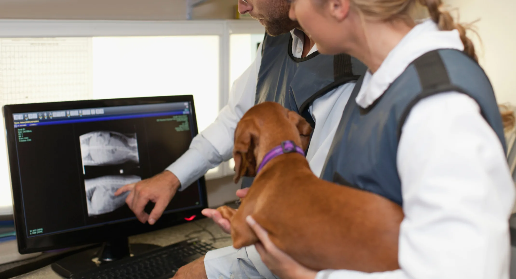
X-rays (i.e., radiographs) are usually the first imaging studies we employ to diagnose your pet’s medical issues. X-rays generate an image when radiation is passed through a particular body part. With digital radiography, the image is captured on a digital recording device and almost immediately displayed on a computer screen. Dense tissue, such as bone, does not allow much radiation passage, resulting in an image that appears white. Less dense structures, such as air-filled lungs, allow most of the radiation to pass through, so this area appears black in the image. Your pet will need to remain still during this procedure and may require sedation. Situations in which X-rays help us diagnose your pet’s problem include:
Bony Abnormalities
— Bones are easily evaluated on X-rays, and conditions such as fractured bones, dislocated joints, arthritis, and bone deterioration can be appreciated.
Dental Disease
— Tooth roots and associated bony structures can be evaluated for periodontal disease.
Organ Abnormalities
— The size and shape of your pet’s organs can be evaluated using X-rays.
Foreign Objects
— If you suspect that your pet has swallowed a foreign object, an X-ray can usually determine where the object is located in your pet’s gastrointestinal tract before they are taken to surgery. X-rays may not show items made of string, fabric, or plastic, however, other imaging modalities may be required.
Tumors
— Some internal tumors can be found on X-ray.
Bladder Stones
— Crystallization inside your pet’s bladder can be seen on X-ray.
Pregnancy
— If your pet is pregnant, the individual skeletons of the developing fetuses can be observed on X-ray.
What an Ultrasound Tells Us About Your Pet
An ultrasound uses a harmless, high-frequency sound beam to image an area, and is usually used as a complement to X-rays. Typically, minimal restraint or sedation is required to perform this diagnostic procedure. Ultrasounds are used to evaluate your pet for a variety of issues, including:
Heart Conditions
— An ultrasound to evaluate your pet’s heart is called an echocardiogram and can help determine how your pet’s heart is functioning. If they have a murmur, the echocardiogram can visualize what structures are causing the problem.
Foreign Objects
— If your pet has ingested a foreign body that cannot be observed on X-ray, an ultrasound may show the object.
Organ Evaluation
— Your pet’s organs can be better evaluated on ultrasound. If something appears suspicious on an X-ray, an ultrasound reveals a more detailed look at the organ in question.
Soft Tissue Injuries
— Ultrasound can be used to assess ligament and tendon injuries.
Eyes
— Eye structures can be evaluated using ultrasound, especially if a condition, such as cataracts, prevents visualization through the lens.
Pregnancy
— Fetal viability can be assessed using ultrasound.
Biopsy Collection
— Ultrasound can be used to guide biopsy collection, to ensure pinpoint accuracy.
Emergency Situations
— An ultrasound can gather valuable information quickly during an emergency situation and can help diagnose conditions such as internal hemorrhage, allowing for prompt treatment.
Endocrine Disease
— The thyroid and parathyroid glands can be evaluated using ultrasound to help diagnose conditions such as hyperthyroidism or hypothyroidism.
Some specialized ultrasounds, including echocardiograms, and eye and ligament evaluations, may require referral to a specialist.
What a Computed Tomography Tells Us About Your Pet
Computed tomography (CT) uses a narrow beam of X-ray radiation aimed at a pet and quickly rotated around their body, producing signals that generate cross-sectional images that contain much more detail than digital X-ray. CT is invaluable for evaluating lesions found during a clinical exam or with preliminary imaging methods. Your pet will need to be anesthetized for this procedure, to ensure the images are clear. Situations in which CT will be employed to diagnose your pet include:
Pre-surgical Planning
— If your pet has a tumor that needs surgical removal, a CT scan helps determine the surgical approach by revealing the tumor’s precise location and relationship to neighboring structures. This knowledge will also minimize surgery time.
Check for Metastasis
— CT scans can be used to assess organs, such as the lungs, for the spread (i.e., metastasis) of malignant tumors.
Assess Trauma Patients
— In severe trauma cases, such as your pet being hit by a car, CT scans can quickly assess any internal damage.
Orthopedic Abnormalities
— CT scans can provide a detailed view of bones and joints, and offer a clearer image of the problem.
Cranial Abnormalities
— CT scan is far superior to X-rays for imaging intricate head anatomy. Studies include:
Brain
— CT scans are used to evaluate the brain for tumors, internal bleeding, trauma, infections, and fluid accumulation.
Nasal Cavity and Paranasal Sinuses
— Abnormalities, such as tumors, inside these structures can be better assessed using CT scans because the cross-sectional view prevents superimposed structures from confusing the image.
Dental Exam
— Tooth roots can be evaluated using a CT scan if your pet is affected by periodontal disease.
Ears
— Structures inside the ear can be easily visualized with CT scans, but not X-rays or ultrasound.
Diagnostic imaging helps us to determine what is wrong with your pet when they cannot speak for themselves. If your family veterinarian believes your pet requires imaging to help diagnose a medical issue, do not hesitate to contact our team at VVC.
